How to Achieve the Optimal Endpoint Response for Laser Freckle Removal

Laser is the abbreviation for light amplification by stimulated emission of radiation (Laser). Different media produce different wavelengths of laser, corresponding to different target color groups (melanin, hemoglobin, water, etc.) with absorption peaks and penetration depths in the skin. The selection of wavelength and energy is important for the clinical use of laser equipment to treat pigmentary diseases. In addition, it is necessary to consider the immediate endpoint response of the treatment to help determine the efficacy and prognosis of the treatment.
1. Freckles
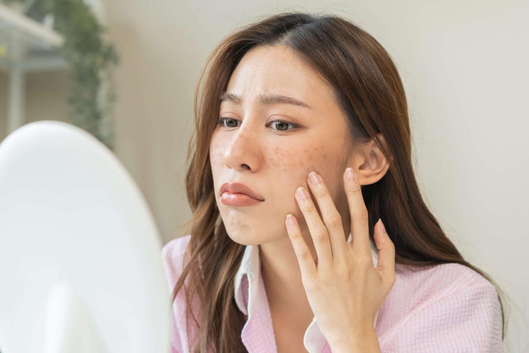
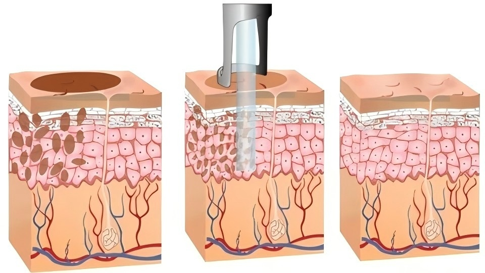
Freckles are brown punctate pigmentation spots commonly found on the face, which are autosomal dominant genetic diseases that can be promoted and exacerbated by sun exposure. Pathological examination shows that there are a large number of melanin granules on the epidermis of the freckle lesion site, but the number of melanocytes has not significantly increased.
1.1 Treatment Methods
Freckles are often treated with laser or intense pulsed light in clinical practice, which generally have good therapeutic effects, but there is a possibility of recurrence in the later stage. Postoperative sun protection can reduce or delay recurrence. The shorter the wavelength, the higher the melanin absorption coefficient, but short wavelength lasers penetrate the skin too shallowly. In clinical practice, lasers with wavelengths such as 532nm, 694nm, 730nm, and 755nm are commonly used for treatment. When selecting strong pulsed light for treatment, light in the 500-600nm wavelength range is preferred.
1.2 Endpoint Reaction
Different devices have different endpoint reactions. For nanosecond level lasers, such as Q-switched Nd: YAG 532nm lasers, the endpoint reaction is generally dark gray or slightly frosted, and it is best not to have severe frosting or even skin splashing. For picosecond lasers, due to the additional light pressure effect, melanin particles can be shattered into smaller fragments, and the endpoint response of treatment is generally lighter than that of nanosecond laser devices, with a dark gray change.
When using strong pulsed light to treat freckles, obvious pigmentation can usually be seen immediately after treatment (darkening of the color spot), and the skin around the color spot is slightly reddish.
1.3 Adverse Reactions
If the energy during treatment is high, adverse reactions such as blisters may occur after surgery, which may result in darkening, depigmentation, and even scarring. The postoperative treatment area usually forms scabs, and patients should be advised to avoid rubbing the treatment area to prevent the scab from falling off prematurely and increasing the risk of discoloration.
After treatment, attention should be paid to strengthening sun protection to reduce the risk of postoperative pigmentation and recurrence. If there is a decrease in pigmentation after surgery, it can generally recover on its own over time.
1.4 Treatment Recommendations
When treating freckles for the first time, try to achieve sufficient endpoint response. If the energy density is not sufficient, the color of the freckles will become lighter after one treatment, and the absorption selectivity of laser in subsequent treatments may be weak, making treatment more difficult. Treatment should pay attention to preventing color fading.
For people at high risk of color fading, local area treatment can be chosen to observe possible color fading reactions. For patients with freckles accompanied by melasma, the condition of melasma can be stabilized first, and then freckle treatment can be carried out, which can be combined with oral medication and other treatments to prevent and reduce pigmentation reactions.

2. Brown Spots
Melasma is a symmetrical distribution of light brown to dark brown pigment spots mainly on the cheekbones and cheeks. It can also be seen on the forehead, cheeks, upper lip, etc. It is common in women of childbearing age, especially during pregnancy, and can also occur in men. The pathogenesis of melasma is still unclear and may be related to pregnancy, oral contraceptives, certain medications, cosmetics, liver disease, genetics, and ultraviolet radiation.
Pathological examination shows melanin deposition in the skin lesions of melasma, mainly in the basal layer and above, with visible damage to the basement membrane, and melanocytes in the dermis layer. The number of melanocytes is normal, but dendrites are significantly enlarged and melanin levels are increased.
2.1 Treatment Methods
The first-line treatment for melasma is systemic or topical medication, which can be combined with chemical exfoliation and phototherapy to reduce melanin production and promote melanin metabolism. The patient’s melanocyte function is relatively hyperactive, and physical and chemical therapy should be mainly mild. The energy of phototherapy should not be too high, and longer wavelengths are generally selected, such as 755nm and 1064nm. Large spot low-energy toning mode is generally used, with an interval of about 2 weeks.
2.2 Endpoint Reaction
The skin lesion area is slightly red or shows no significant changes. Try to avoid using explosive scab removal mode to treat melasma skin lesions, and avoid changes such as dark gray or white, purpura, etc.
2.3 Adverse Reactions
When the energy of laser treatment is too high, patients with melasma are at a higher risk of color darkening, and the color darkening subsides slowly, which may lead to serious consequences such as pigment incontinence.
2.4 Treatment Recommendations
During the progression of melasma, oral medication should be the main treatment method to avoid irritating treatment in the affected area. During stable treatment, laser energy should not be too high, and the main effect should be on light modulation to stabilize pigment cell function. Chemical exfoliation can be combined to promote melanin metabolism. Due to the pathological manifestation of photoaging in melasma, combined treatment with dot matrix microneedle radiofrequency can improve the inflammation and aging microenvironment of melasma.
3. Seborrheic Keratosis
Seborrheic keratosis, commonly known as age spots, is a benign epidermal tumor caused by delayed maturation of keratinocytes. The etiology is unknown and is related to age and sun exposure. The skin lesions are pale yellow to dark brown patches or flat papules or verrucous papules. The pathology is characterized by hyperkeratosis, thickening of the spinous layer, and papillary hyperplasia.
3.1 Treatment Methods
Skin lesions with obvious pigment deposition and mild hyperplasia can be treated with laser or intense pulsed light similar to freckles. If the hyperplasia is thick, exfoliative laser such as CO laser or bait laser can be selected for grinding treatment in the same treatment, and then Q-switch laser can be used for pigment treatment.
3.2 Endpoint Reaction
The treatment methods vary depending on the thickness. The treatment for thinner hyperplasia is similar to freckles, and the endpoint reaction may be slightly heavier than freckle treatment, with visible frost white. When grinding treatment for thick hyperplasia, the treatment depth should preserve the bottom layer of skin lesions and avoid damage and bleeding as much as possible. Q-switch laser treatment can be selected for the bottom layer, and the endpoint reaction can be visible as frost white.
3.3 Adverse Reactions
When the treatment energy is high or the depth is too deep, there may be damage, blisters, purpura, and subsequent adverse reactions such as heavy pigmentation, depigmentation, or scars.
3.4 Treatment Recommendations
Control the energy and depth of treatment to avoid scar formation.p
